Positron Emission Tomography (PET) is a medical scanning technique which is used to investigate normal and abnormal processes in the body. The images produced by a PET scanner reflect how the tissue is functioning at the molecular level. Most other types of scanner, e.g. Computer Tomography (CT) or Magnetic Resonance Imaging (MRI) shows images that reflect the anatomy of the body, whereas a PET image may reflect, for example, the amount of oxygen that is reaching a tumour or whether a chemotherapy drug has acted upon a particular cellular pathway. For this reason, PET is generally classified as a 'molecular imaging' technique.
PET works by injecting a very small amount of a radiolabelled compound into the body, usually intravenously. The compound is selected to be taken up by a particular process. For example, a radiolabelled glucose analogue (fluorodeoxyglucose or FDG) is taken up by tissues in proportion to its rate of metabolism, and in particular by most tumours. Radiation emitted from the radiotracer escapes from the body and is detected by the PET scanner, which then constructs a 3D image of the distribution of the radiotracer. Regions that have accumulated tracer (e.g. a tumour) show up as 'hot spots' on the PET image. The radioactive label has a half-life of just a couple of minutes to a couple of hours and so rapidly disappears from the body. The radiation dose associated with a PET scan is roughly equivalent to several years natural background radiation.
FDG is the most widely used PET radiopharmaceutical. The scanning session starts with injection of FDG, the patient must then remain still for ~60 minutes for the tracer to distribute within the body. The patient then lies in the PET scanner for ~20 minutes whilst the image is obtained.
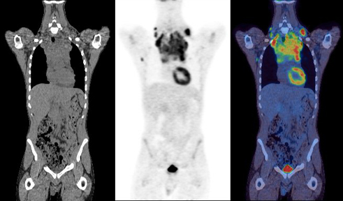
A. Pre-treatment Images (CT, PET and fused PET-CT image, from left to right)
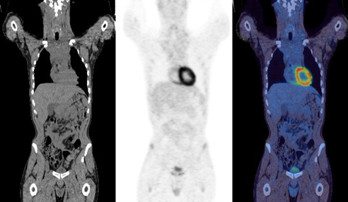
B. Post-treatment Images (CT, PET and fused PET-CT image, from left to right)
'Positron Emission Tomography' refers to the fact that the radioactive label which allows the injected compound to be imaged, emits a particle called a positron. The idea of imaging positron labelled compounds goes back to the early 1950s. One reason that PET has taken so long to become widely available is that it is complex and hence expensive. In addition to the PET scanner, a cyclotron is required to produce the positron emitting radionuclide, and a radiochemistry laboratory is needed to attach this radioactive tag (e.g. 18F) to the relevant compound (e.g. glucose). Therefore, both routine and research PET activities are truly multidisciplinary, requiring technical expertise from chemists, physicists, computer experts, physicians, pharmacists, biologists, and radiographers.
Nearly all PET scanners in use today have a CT scanner incorporated into the same gantry. The CT scanner uses x-rays to obtain a high resolution anatomical image of the body. A CT scan, which only takes ~30 seconds, is performed immediately prior to the PET scan. The anatomical CT scan allows 'hot spots' and other features seen on the PET image to be located within the body.